Outcrop Modal Analysis,
a Petrographic Application of Digital Image
Processing Technology
Tim Allen, Geology Dept., Keene State College, Keene, NH 03435-2001, 603-358-2571, tallen@keene.edu
Introduction
Quantitative determination of the modes of components in rocks is an important petrographic task, providing necessary data to understand the petrologic history of rocks. Commonly this is done by point counting of thin sections as viewed through a petrographic microscope. For very coarse-grained or heterogeneous rocks, the thin section no longer provides a representative sample of the rock. In some cases, it may even be necessary to count modes at the outcrop. In a recent study of pelitic migmatite gneisses (see Figure 1a and Figure 1b) in the White Mountains of New Hampshire (Allen, 1992), I had to determine the modes of leucosome (the light colored, granitic part), melanosomes (the dark colored, restitic part), and mesosomes (the remainder, of intermediate color and composition). I used two techniques to determine these modes: point counting with a wire mesh on the outcrop, and classification of digital images obtained by scanning photographs of the outcrop surfaces (Allen, 1994). This second technique is a rather novel application of remote sensing technology, so I will describe the method and compare it with the point counting.
Point Counting
I used a 30" by 30" piece of 1/2" wire mesh -- "hardware cloth" available at any hardware store -- to provide a grid with which to count points. This mesh covers an area of about 0.58 m2 and includes approximately 3600 points. The location of areas to count at the outcrop were chosen at random, except that a smooth contiguous surface was preferred. These surfaces were generally glacially polished or water worn, although other surfaces were also counted. The modal results from point counting are given in Table 1.
Imaging
I took photographs of the same areas that were point counted, using standard black and white negative film (ISO 100) in a 35mm SLR camera, with 50mm normal focal length and 28mm wide-angle lenses. Every effort was made to keep the film plane parallel to the outcrop surface being imaged to limit perspective distortions, although this was not always possible. The wide angle lens allows a shorter camera-to-subject distance for the same image area, which is an advantage when trying to keep the film plane parallel to the outcrop surface while photographing down onto a horizontal outcrop surface, or on an outcrop on a side hill.
I took the photographs on an overcast day, which provides soft, even, flat lighting. Direct sunlight striking the outcrop surface at an angle will enhance any irregularities in the surface, as these will cast shadows. In addition, it is possible that the contrast in a scene under bright sunlight might exceed the tonal range of the film. The film was exposed according to the camera's exposure meter; and was processed by standard methods at a local photo-finisher.
The standard size black and white prints obtained from the photo-finisher were digitized using a flat-bed scanner capable of creating 8-bit grayscale images. The scanner measured the brightness or gray level of each pixel in the photograph, and assigned to that pixel an integer value in the range from 0 (black) to 255 (white). This is the range of integer numbers representable by a single byte, which is a standard method of representing digital numbers. Minimal adjustments to brightness and contrast were made with the scanner software during scanning of the first image to produce a visual match between the digital image on the screen and the original photograph. It appears that the scanner software may also have applied an automatic histogram "stretch" to produce digital images with a normal distribution, using the full range of possible brightness values (0 to 255).
The photographs were scanned at a resolution of 75 pixels per inch, yielding approximately 50,000 pixels within the area of interest in each photograph (the 0.58 m2 area that was manually point counted). Higher scanning resolutions are possible, but these would also exponentially increase the disk storage space required for each image. The resolution used was quite adequate. The scanned images are Figure 1a and 1b.
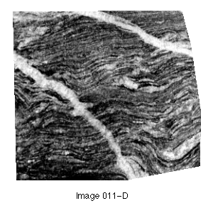
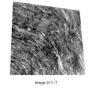
Figures 1a (left) and 1b (right)
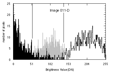
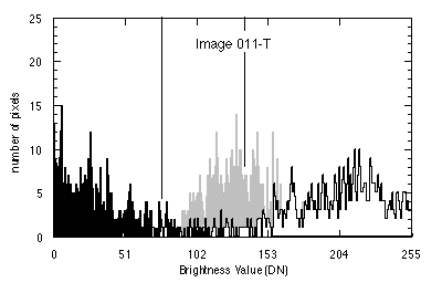
Histograms, showing the number of pixels in the scene at each brightness level, are shown in Figure 2a and Figure 2b. The area of Figure 1a includes two pegmatite veins. This veins were selected and cut out of the digital image prior to further processing and these pixels are not included in the histogram for this scene. The presence of these bright veins in the photograph during scanning, however, did affect the distribution of brightness values in the remainder of the scene. This is indicated by the slightly skewed histogram for this image (not including the veins; Fig. 2a).
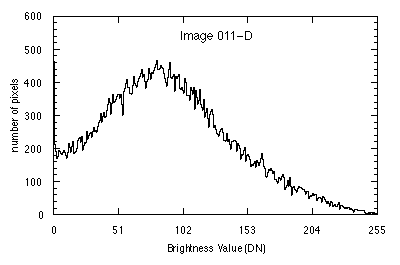
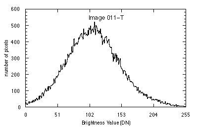
Figures 2a (left) and 2b (right): Histograms of number
of pixels for each brightness value (Digital Number or DN), for the
scenes shown in Figures 1a and 1b respectively. The histogram in Figure 2a does not inclcude pixels from
the cross-cutting pegmatite veins.
Classification
In this study of migmatites, I was interested in three categories or classes: leucosomes, melanosomes, and mesosomes. In order to classify the digital images, I needed to establish criteria with which each pixel could be put into one of these classes. To do this, I measured the pixel values in "training sites" for each of the three classes. These training sites were picked by visual inspection of the images, and the pixels in the training area were selected by outlining the area using a cursor. A histogram of pixel values in the selected areas were recorded, along with other measurements. These are presented in Table 2. The areas of training sites for each of the classes represented approximately 1% of the total scene area.
Figures 3a (left) and 3b (right): Histogram of number of pixels for each brightness value (Digital Number or
DN), for leucosome (white), mesosome (gray), and melanosome (black) training
sites selected from the images in Figures 1a and 1b respectively. The number of pixels included
in each training site is given in Table 2. Vertical
lines show the threshold values used for classification (Table 2). The resulting classified images are shown in Figures 4a and 4b respectively.
The histograms from the training areas are shown in Figure 3a and Figure 3b. As can be seen, there are overlaps in the brightness values of leucosome, mesosome, and melanosome. Thus classification required determining "cut-off" or threshold values for the boundaries between the three classes. The thresholds used in this study are given in Table 2. The upper threshold for the melanosome class was taken to be the mean plus 1.5 standard deviations, as determined from the melanosome training sites. The lower threshold for the leucosome class was taken to be the mean minus 1.5 standard deviations, as determined from the leucosome training sites. The choice of 1.5 standard deviations was fairly arbitrary, but is representative of the boundaries between melanosome, mesosome, and leucosome (Fig. 3b). The long tails and substantial overlap of the histograms for the training sites in Figure 1b (Fig. 3b) indicate that the areas chosen as training sites were extremely heterogeneous. This suggests that sites were not chosen carefully enough or that leucosome and melanosome are intermingled at a finer scale than the size of the chosen training areas. For Figure 1a, it was necessary to adjust these values due to the skewing of the distribution of brightness values for the whole image (Fig. 2a). Thus for this image, the upper threshold for the melanosome class was taken to be the mean plus 1.0 standard deviations, as determined from the melanosome training sites. The lower threshold for the leucosome class was taken to be the mean minus 2.0 standard deviations, as determined from the leucosome training sites. These values are also representative of the boundaries between leucosome, mesosome, and melanosome (Fig. 3a).
The classified images are shown in Figures 4a and 4b, where white represents the leucosomes, gray--the mesosomes, and black--the melanosomes. The modes determined by these classifications are given in Table 1.
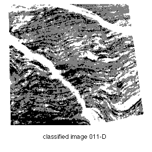
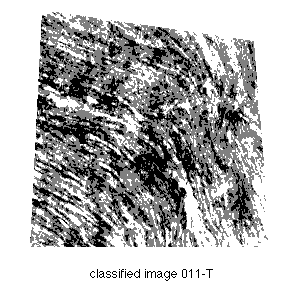
Figures 4a (left) and 4b (right): Classified images using the respecitve threshold values depicted in Figures 3a and 3b.
The histogram for the original images (Figures 1a and 1b) are shown in Figures 2a and 2b. Statistics
for the whole scene and the training sites are given in Table
2, and classification results in terms of modes of Leucosome, Mesosome and Melanosome are given in Table 1.
TABLE 1
                Leucosome   Melanosome  Mesosome

011-D (digital)  17.7        24.7        57.6
011-D (manual)   30.0        32.0        38.0
(Figure 1a)

011-T (digital)  24.8        24.0        51.2
011-T (manual)   20.0        33.0        47.0
(Figure 1b)

036-T           25.7
036-D           31.5
038-T           13.4
038-D           29.3
039-1-T         25.3
039-1-D         26.2
039-4-T         22.8
039-4-D         22.1
Table 1: Modal analyses of migmatite outcrops, giving percentage of total
surface area (equivalent to volume) occupied by leucosome, mesosome, and
melanosome.
TABLE 2
            # of Pixels     Mean    s. d.   Mode    Min     Max     T-hold
Figure 1a (Image 011-D)
Whole Scene   54452          90.6   50.9      0 (?)  0      255
    Training sites (Figure 3a):
    Leucosome   824         201.3   31.1    219     73      255     139
    Mesosome    582         106.2   29.0    111     39      195
    Melanosome  528          24.9   27.0      0      0      173     52

Figure 1b (Image 011-T)
Whole Scene   49318         108.2   42.7    109      0      254
    Training sites (Figure 3b)
    Leucosome   502         195.8   39.8    217     49      254     136
    Mesosome    470         133.5   25.2    130     52      204
    Melanosome  352          33.3   29.2      5      0      151      77
Table 2: Image classification statistics: mean, standard deviation, mode, minimum and
maximum brightness values for the numbers of pixels measured. The T-hold
column shows the threshold values used to distinguish between Leucosome,
Mesosome, and Melanosome classes in classifying the images (Fig. 5 and Fig. 6;
classification results given in Table 1).
Discussion
The digital classification method relies entirely on relative differences in brightness of the classes to be measured. Manual point counting, on the other hand, allows the geologist to use additional information such as mineralogy and texture to determine in which class each point belongs. In addition, while carrying out the point count, the geologist is afforded an intimate look at the textures of the rock, an opportunity that might otherwise be missed. The digital classification allows a much finer resolution, and the possibility of measuring a much larger surface area than would be feasible by point counting. In addition, the digital classification applies the classification criteria uniformly across the whole image scene, whereas during manual point counting, the person counting may subjectively change the criteria as the count drags on. This type of subjectivity is particularly a problem for loosely (and somewhat subjectively) defined classes such as the leucosome, melanosome, and mesosome of migmatite outcrops. (For thin section point counting of mineral modes, quartz and biotite are clearly defined and easily distinguished.) This comparison (Table 1) of these two methods suggests that manual point counting, or at least close inspection of the outcrop, may provide necessary "ground truth", but that the application of remote sensing techniques as used here may be an effective method of enhancing and extending outcrop-scale petrography.
Equipment and Software
The photographs were taken with an Olympus OM-1 35 mm single-lens reflex camera, with Olympus-Zuiko 50 mm, f1.4 and 28 mm, f2.8 lenses, on Kodak Plus-X (100 ISO) panchromatic film. The images were scanned with a La Cie SilverScanner, controlled by a plug-in software module for the Adobe Photoshop program. The image processing was originally done on an Apple Macintosh II computer with an Apple 13" Color monitor driven by a 8-bit color monitor card, using the public-domain program, NIH Image, version 1.44, developed by Wayne Rasband of the National Institutes of Health.
References Cited
Allen, T., 1994, Outcrop Modal Analysis, a Petrographic Application of Digital Image Processing Technology (abstract), GSA Abstracts with Program, v. 26, n. 3, p. 2.
September 1994
tallen@keene.edu